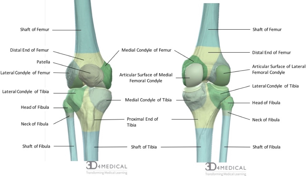
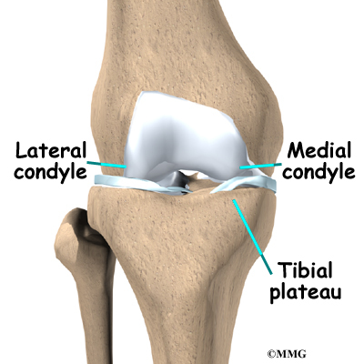
The femoral condyles are the two rounded prominences at the end of the femur. The femoral condyles articulate or contact with the tibia and on the medial side this is in the medial tibial plateau and the medial meniscus and on the outside of the knee is known as the lateral tibial plateau in the.
Bones Advanced Anatomy 2nd Ed
The posterior and inferior surfaces articulate with the tibia and menisci of the knee while the anterior surface articulates with the.
. The medial condyle is one of the two projections on the lower extremity of femur the other being the lateral condyle. The tuberosity of the tibia or tibial tuberosity or tibial tubercle is a large oblong elevation on the proximal anterior aspect of the tibia just below where the anterior surfaces of the lateral and medial tibial condyles end. In the 155 varus knees the radius of the lateral condyle was an average of 01 mm larger than that of the medial condyle p 0003.
Interface of the medial and lateral femoral condyles. The intercondylar fossa of the femur is located 50 between the lateral and medial epicondyles of the femur between the greater and lesser trochanter of the femur between the lateral and medial condyles of the femur between the lateral and medial condyles of the humerus between the lateral and medial plateau of the tibia. The radius of a condyle was the average of the radii on four adjacent images that showed the femoral condyle with the largest curvature.
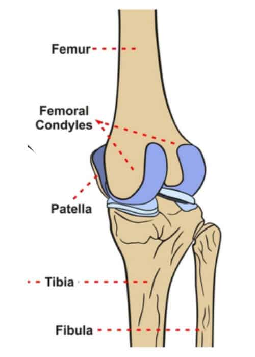
There is a significant difference in articular cartilage thickness between the medial and lateral posterior femoral condyles in patients undergoing unicompartmental knee arthroplasty. A diagram of the knee showing the medial femoral condyle where the medial collateral ligament attaches to the femur. They are called the medial and the lateral femoral condyle respectively.
There are two condyles on each leg known as the medial and lateral femoral condyles. D 0336 WDC -0097 WMC -0153 DLC 0372 DIN - 20912. The femur is the bone commonly known as the thigh bone.
The articular cartilage thickness on the medial posterior femoral condyle was 3 mm 1 mm mean standard deviation and 1 mm 1 mm on the lateral side p-value. The paired femoral condyles are situated to either side of the patella or kneecap. Anatomical terms of bone.
Meniscus Tear Medial Medial Meniscal Tear Medial meniscus and ACL tear happens when a flexed knee is twisted inwards medially with the foot firmly planted on the ground. The p-value for statistical significance was set at 005. If there is a fracture break in part of the condyle this is known as a fracture of the femoral condyle.
The medial and lateral condyles are the epiphyseal ends of the femur that articulate with the tibia and the patella. They are the pair of rounded eminences that form the. The medial condyle is one of the two projections on the lower extremity of femur the other being the lateral condyle.
The medial condyle is larger than the lateral outer condyle due to more weight bearing caused by the centre of mass being medial to the knee. Mean medial condyle BMDs medial versus lateral condyle BMD ratios and visual analog scale VAS pain in both the femur and the tibia were higher in the obliteration group compared with the narrowing group P 0001 for all. A femoral condyle is the ball-shape located at the end of the femur thigh bone.
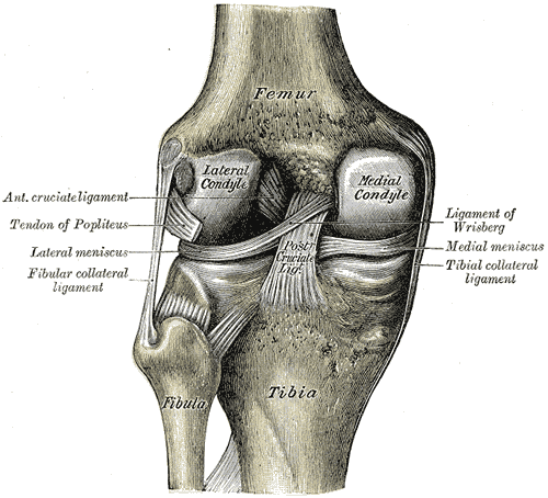
It is the most commonly injured meniscus. Both condyles are smooth and rounded posteriorly to allow for motion and level out inferiorly allowing for articulation with the tibia. They are separated by the deep intercondylar fossa proximally bounded by the horizontal intercondylar line.
Any abnormal surface structure or cartilage damage can lead to cartilage breakdown and arthritis loss of cartilage padding. 4 rows In other words the lateral surface of the medial condyle the medial wall of the. On each condyle is a smaller epicondyle which serve as the point of attachment for the collateral ligaments the medial collateral MCL and the lateral collateral ligaments LCL.
What movement does the MCL prevent. They are called the medial and the lateral femoral condyle respectively. A significant positive correlation was observed between the femoral and tibial condyles in the following parameters.
The motions of the condyles include rocking gliding and rotating. The medial condyle is one of the two projections on the lower extremity of femur the other being the lateral condyle. The motions of the condyles include rocking gliding and rotating.
Asked Aug 23 2019 in Health Biomechanics by Livaco. Learn more about the femur in this. The medial condyle is one of the two projections on the lower extremity of femur the other being the lateral condyle.
The medial meniscus is a C shaped immobile structure covering nearly 50 of the medial tibial plateau. The medial femoral condyles are the bony protrusions on the inside edge of the bottom of the femur bone in each thigh. The most accurate equation used width of the medial and lateral condyles WDC with of the medial condyle WMC depth of the lateral condyle DLC and depth of the intercondylar notch DIN 941 and is as follows.
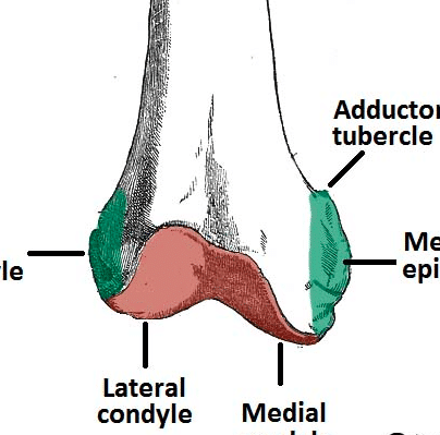
While both bones feature a medial and lateral condyle with the lateral condyle on the other side of the knee the medial condyle is the larger prominence because more weight is transferred across the inside aspect of the knee joint. The medial femoral condyle is located on the inside part of the knee whereas the lateral femoral condyle which is bigger is located on the outside part of the knee. At the end of the medial supracondylar line is a tubercle called the adductor tubercle.
The medial condyle is larger than the lateral outer condyle due to more weight bearing caused by the centre of mass being medial to the knee. In between the medial and lateral femoral condyles is the intercondylar fossa. The femur articulates with the medial and lateral condyles of the tibia.
Cartilage thickness of the lateral posterior femoral condyle and medial posterior femoral condyle was recorded and expressed in millimeters mm. On the posterior surface of the condyle the linea aspera a ridge with two lips. The medial condyle is convex in shape with a width around 2532 mm while the lateral.
Bones of the Knee Joint The femoral condyles are the two rounded prominences at the end of the femur. Where is the medial femoral condyle of the knee. Differences in articular cartilage thickness between the means of these two groups medial and lateral were evaluated using Students t-test.
Medial and lateral condyles rounded areas at the end of the femur. Similar to the articular surface of the patella the trochlear surface is divided into medial and lateral facets the lateral facet being larger and extending more proximally and anteriorly than its medial counterpart Figure 22-2. The distal end of the femur is characterised by the presence of the medial and lateral condyles which articulate with the tibia and patella to form the knee joint.
The femoral condyles form the trochlear groove that provides the articulating surface of the femur. The lateral condyle is called the capitulum and the medial condyle is called the trochlea. The distal end of the humerus forms two condyles.
The medial and lateral condyles of the tibia articulate with the. The medial condyle is larger than the lateral outer condyle due to more weight bearing caused by the centre of mass being medial to the knee. Palpable as a hard rounded bump to the inside of either knee joint they are one of two condyles at the.
The medial and lateral condyles form the proximal part of the body of femur and articulate with the proximal part of tibia to form the femorotibial joint.
Femoral Condyle Articular Cartilage Injury Minneapolis St Paul Edina Eagan Mn
2
Medial Condyle Of Femur Wikipedia
Distal Femur Fracture Teachmesurgery

Bones Specialist Knee Surgeon In Manchester Professor Sanjiv Jari
Orif Lag Screw For Lateral Medial Femoral Epicondyle Fracture

Femur An Overview Sciencedirect Topics
Eorthopod Patient Education
0 comments
Post a Comment